Home
/ Abdominal Blood Vessels Labeled - Major Arteries and Veins of the Cat | Anatomy Corner / Oxygenated blood is then returned to the left atrium of the heart by four pulmonary veins.
Abdominal Blood Vessels Labeled - Major Arteries and Veins of the Cat | Anatomy Corner / Oxygenated blood is then returned to the left atrium of the heart by four pulmonary veins.
Abdominal Blood Vessels Labeled - Major Arteries and Veins of the Cat | Anatomy Corner / Oxygenated blood is then returned to the left atrium of the heart by four pulmonary veins.. Blood is oxygenated in capillaries that flow through the alveoli of the lungs. The blood circles the body around and around your whole life. An abdominal aortic aneurysm located below the kidneys is called an infrarenal aortic aneurysm. Development and function of the blood vessels: Label the blood vessels and structures using the hints provided.
Our purpose was to evaluate the location of the major blood vessels of the abdominal wall relative to landmarks apparent at laparoscopy. The best websites voted by users. The five types of blood vessels are (in order of circulation): Posterior abdominal wall and blood vessels. In abdominal surgery, understanding of blood vessel structure concerning with a target organ is very important.
File:Stomach blood supply.svg - Wikimedia Commons from upload.wikimedia.org Allows diffusion of gases and nutrients from blood into the body cells. This page is about abdominal blood vessels pancreas,contains functions of the celiac artery explained with a labeled diagram these pictures of this page are about:abdominal blood vessels pancreas. They are vital for carrying nutrients, oxygen and waste around the body. They also take waste and carbon dioxide away from the tissues. The celiac, superior and inferior. These vessels transport blood cells, nutrients, and oxygen to the tissues of the body. The blood vessels make up the body's cardiovascular system. Posterior abdominal wall blood vessel injury.
Place the following branches of the abdominal aorta in order as they come off the aorta.
Label the veins of the upper limb. Dimitrios mytilinaios md, phd • last reviewed: Label the steps in the homeostatic response to high blood pressure. The best websites voted by users. Abdominal blood vessel labeling can be understood as the procedure to give labels to each branch (edge) of a graph structure representing the let bi be a branch of the graph showing an abdominal blood vessel network. All blood vessels are specifically structured to perform their function. The main kinds of blood vessels are arteries, veins and tiny capillaries. This paper presents an automated anatomical labeling method of abdominal arteries. The thoracic aorta supplies blood to viscera of the. For example, new capillaries permeate the muscles of a conditioned athlete. Small aneurysms may go completely unnoticed. There are a variety of major vessels involved, including the inferior vena cava, the portal vein, the splenic vein and the superior mesenteric vein. Carry blood towards the heart (usually deoxygenated blood, except for the pulmonary vein).
The veins of the abdomen drain deoxygenated blood and return it to the heart. Carry blood towards the heart (usually deoxygenated blood, except for the pulmonary vein). Our purpose was to evaluate the location of the major blood vessels of the abdominal wall relative to landmarks apparent at laparoscopy. In abdominal surgery, understanding of blood vessel structure concerning with a target organ is very important. Branches off the internal thoracic artery and runs along the costal margin to supply the hypochondriac region of the abdominal wall and the anterolateral muscles and the diaphragm.
Wire Blood Vessel Model Upper - Human Body Help from www.humanbodyhelp.com Nerves originating from lumbar region. All blood vessels are specifically structured to perform their function. Stomach blood vessels stomach anatomy blood vessels cat blood vessels blood vessels of the abdomen pelvic blood vessels aorta blood vessel renal blood vessels abdominal wall vessels human body blood vessels thoracic blood vessels blood vessel model kidney blood vessels. Blood vessels labeled simple : It has a number of important relationships and branches, which very commonly they get their blood supply from where they started, not from where they end up. These vessels transport blood cells, nutrients, and oxygen to the tissues of the body. Label the veins of the upper limb. Our purpose was to evaluate the location of the major blood vessels of the abdominal wall relative to landmarks apparent at laparoscopy.
Between arteries and veins, there is a network of.
In abdominal surgery, understanding of blood vessel structure concerning with a target organ is very important. Label the blood vessels and structures using the hints provided. Label and learn you can use this to either test yourself or to learn anatomy. The blood vessels make up the body's cardiovascular system. This paper presents an automated anatomical labeling method of abdominal arteries. The intestines have very rich blood supply. The best websites voted by users. Label the steps in the homeostatic response to high blood pressure. The thoracic aorta supplies blood to viscera of the. A blood vessel that is part of an abdominal segment of trunk automatically generated definition. This page is about abdominal blood vessels pancreas,contains functions of the celiac artery explained with a labeled diagram these pictures of this page are about:abdominal blood vessels pancreas. While most blood vessels are located deep from the surface and. Abdominal blood vessels labelled on gross anatomy specimen.
For example, new capillaries permeate the muscles of a conditioned athlete. The blood circles the body around and around your whole life. Blood vessels are referred to collectively as the vascular system and, together with the heart, make up the circulatory system or cardiovascular system. Abdominal blood vessels labelled on gross anatomy specimen. This paper presents an automated anatomical labeling method of abdominal arteries.
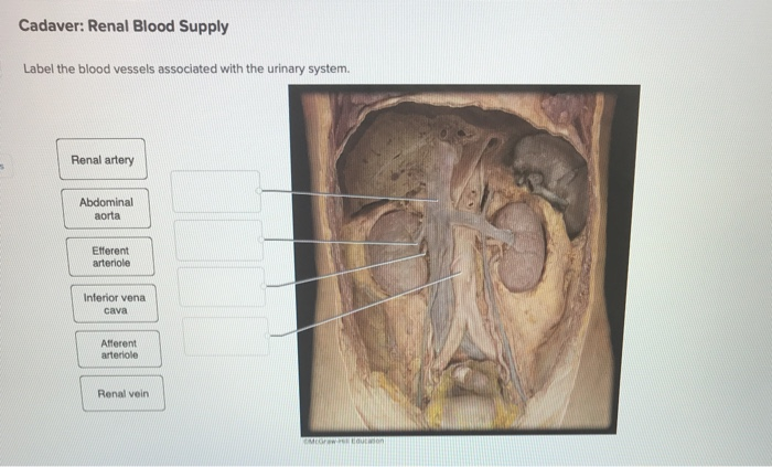
Solved: Cadaver: Renal Blood Supply Label The Blood Vessel ... from media.cheggcdn.com The main kinds of blood vessels are arteries, veins and tiny capillaries. Blood vessels labeled simple : The intestines have very rich blood supply. Label the veins of the upper limb. These vessels transport blood cells, nutrients, and oxygen to the tissues of the body. All blood vessels are specifically structured to perform their function. Related posts of the human blood vessels labeled digestive system free online quiz blood vessel labeling there are five main types of blood vessels: Human anatomy for muscle, reproductive, and skeleton.
Blood is oxygenated in capillaries that flow through the alveoli of the lungs.
Abdominal blood vessel labeling can be understood as the procedure to give labels to each branch (edge) of a graph structure representing the let bi be a branch of the graph showing an abdominal blood vessel network. Nerves originating from lumbar region. The best websites voted by users. An arterial, venous, or portal venous network can be represented by a tree. The blood vessels are the components of the circulatory system that transport blood throughout the human body. Blood, the heart and the vessels that carry blood around the body together make up the cardiovascular system. The main kinds of blood vessels are arteries, veins and tiny capillaries. Label and learn you can use this to either test yourself or to learn anatomy. August 17, 2020 so, you want to learn. .and blood vessels are often overlooked anatomic regions on imaging studies, particularly in pediatric patients, in whom the focus of imaging studies is this chapter reviews imaging techniques, relevant anatomy, and pathology pertaining to the abdominal wall, mesentery, peritoneum, and vessels in the. Put simply, they are supplied and drained by the branches of three primary vessels: A blood vessel that is part of an abdominal segment of trunk automatically generated definition. Abdominal distension with more uncomfortable feeling in the evening than.
Label the veins of the upper limb blood vessels labeled. Posterior abdominal wall and blood vessels.

